Let’s look into the molecules involved in mitochondria’s electron transport chain and oxidative phosphorylation in more detail.

Present on the cristae (inner mitochondrial membrane) are over forty (40) proteins, fifteen (15) of which are directly involved in electron transport. Most of the proteins involved in this are grouped into three large respiratory enzyme complexes.

1-    NADH dehydrogenase complex. This enzyme removes the hydrogen to make it NAD.

2-    Cytochrome b-c1 complex.  Second receiver of the electron.

3-    Cytochrome oxidase complex. Final receiver that will donate to oxygen.

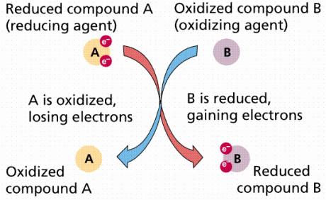
Each enzyme contains metal ions, either iron (Fe) or copper (Cu) in their heme-group (heme- iron or any readily oxidize-able metal ion) that allow passage of electrons through them. Their atomic state changes from being reduced (being more electronegative) when accepting an electron to being oxidized (being less electronegative) when losing an electron.

Redox Reaction. Click on image for credit.

This redox reaction (reduction followed by oxidation) is responsible for the colour change of the protein heme-group in the enzyme complex which is why the word cytochrome (colourful) is used to describe the enzymes. For example, the iron atom changes from ferric (Fe3+) which is yellow to ferrous (Fe2+) which is blue, when it gets reduced (gains electrons to become more electronegative).

During the time that the respiratory enzyme complexes transfer the electrons in the chain, they are also in the same instance pumping protons from the inner mitochondria matrix (with the help of ubiquinone and cytochrome-c) into the intermembrane space. The way this is done is they pick up a proton from the mitochondrial matrix by attracting it to the electron (during the temporary moment when the electrons are in their possession), and then releasing proton into the intermembrane space as the electron is passed on to the next respiratory enzyme complex in the chain.

Allosteric changes creates sequential changes in protein configuration that causes the H+ to enter. Click on image for credit.

The attraction occurs in the form of allosteric (cooperative binding) changes, because the temporary accommodation of the electron within the respiratory enzyme complex creates sequential changes in protein configuration that causes the H+ to enter. Once the electron gets transferred, a similar change in the protein configuration of the enzyme causes the H+ to be released on the other side in the intermembrane space. Think of a newly opened hotel trying to gain guests. It will put out an advertisement of discounted buffet at its dining hall. The people who attend the buffet will then be shown the hotel rooms. Thus, coupling the attraction to the buffet in order to gain prospective clients is similar to coupling the attraction to the electron to attract and pump protons out. The hotel is the enzyme complex, the buffet is the electron, the people attracted to the buffet are the proton, and the hotel rooms (being the intended destination) is the intermembrane space.

Moreover, the active pumping of protons has two major consequences:

  1. It generates a gradient of proton concentration (a pH gradient) across the inner mitochondrial membrane, with the pH about 1 unit higher in the matrix (around pH 8) than in the intermembrane space (close to pH 7).
  2. It generates a membrane potential across the inner mitochondrial membrane, with the inside (matrix side) negative and outside positive as a result of net outflow of protons.

Protein Pump Working due to the built electrochemical gradient. Click on image for credit.

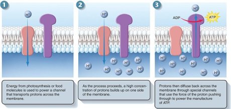
This steep electrochemical proton gradient makes it energetically very favourable for H+ to flow back into the mitochondrial matrix. About 3 protons need to pass through the synthase to make one molecule of ATP (100 molecules are made per second).


ATP synthase is a large, multisubunit protein with an enzymatic portion that looks like a lollipop head and faces inwards and attached, through a thinner multisubunit “stalk”, to a transmembrane proton carrier. The movement of protons down their gradient causes the stalk to spin rapidly within the head inducing it to make ATP.

ATP Synthesis. Credit is due, unfortunately lost the original link. Please inform me to relink if possible.

Btw, the synthesis of ATP is not the only process driven by the gradient, charged molecules (like pyruvate). ADP and Pi are pumped into the matrix from the cytosol while ATP must be moved in the opposite direction. This occurs simultaneously in other carrier proteins present in the cristae which binds these molecules together and couples their transport to the energetically favourable flow of H+ into the mitochondrial matrix (remember cotransport?).
Cotransport. Click on image for credit.

In this post, we will discuss how the components of the mitochondria come together to produce energy or ATP for the cell.

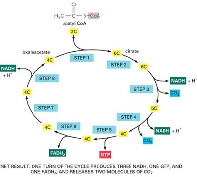
Mitochondria uses, both, pyruvate (mainly glucose plus other sugars) and fatty acids (fat) as fuels. As they enter the mitochondrial matrix, they are converted into acetylCoA (via the enzyme acetyl-coenzyme-A) which is a metabolic intermediate used in the citric acid cycle.

Pyruvate; a component of sugar molecules that enter CAC. Click on image for credit.
Acetyl CoA. It’s many carbon atoms are important for the cycle. Click on image for credit.
Citric Acid Cycle. Click on image for credit.

The above images are for perspective only. Please focus on the text and eye the images as reference. You do not have to memorize everything.

The citric acid cycle converts the carbon atoms in acetylCoA to CO2, which is released from the cell as a waste product.

During this conversion process, the cycle generates high energy electrons carried by carrier molecules NAD+ (Nicotinamide adenine dinucleotide which then becomes NADH) and FAD (flavin adenine dinucleotide which then becomes FADH2). These high energy electrons are then transferred to the inner mitochondrion membrane, where they enter the electron-transport chain.

Electron transport chain on the cristae of the inner mitochondrial membrane now begins.

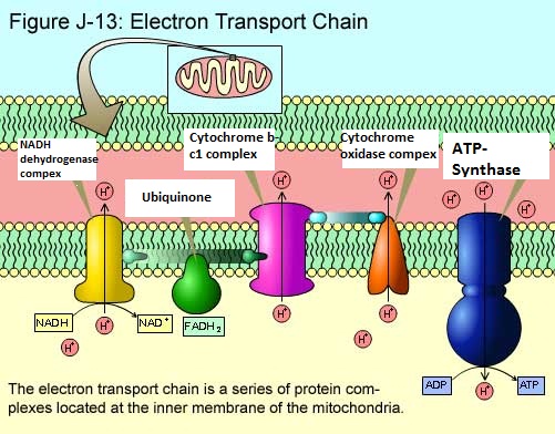
A hydride ion (H) is removed from NADH by the enzyme NADH dehydrogenase complex, and is converted into a proton and two high energy electrons (H -> H+ + 2e). NADH dehydrogenase complex accepts the electrons.

The electrons are then passed along the chain to the second enzyme Cytochrome b-c1 complex with the help of ubiquinone (a small hydrophobic molecule) that picks the electron up from NADH dehydrogenase and delivers it to cytochrome b-c1.

Electron transport chain. Click on image for credit.

The third and final enzyme which receives the electron (this time with the help of cytochrome c) is Cytochrome oxidase complexand as its name indicates, it will finally donate it’s electrons to oxygen.

Actually, four electrons from cytochrome c are added to four protons from the aqueous solution to form two hydrogen molecules (2H2) which then combine with O2 to form water (4e + 4H+ + O2 -> 2H2O). It is here that almost all oxygen we breathe is used, serving as the final receiver for the electrons that NADH donated in the beginning of electron transport chain.

This is also sometimes called the respiratory chain. But what is the point of all this?

Each of the above three respiratory enzyme complexes uses the energy of the electron transfer to take protons from water (present in the aqueous solution H2O dissociating into H+ + OH) and pumps the protons out into the intermembrane space of the mitochondria (explained how later). This creates a build-up of protons in the intermembrane space, building up the electrochemical proton gradient.

Electron transport chain showing clearly the ATP synthase at work. Click on image for credit.

A large inner membrane-bound (keep in mind all this is happening in the mitochondria’s cristae, which is the membrane infolding) enzyme called ATP synthase creates a hydrophilic pathway that allows protons to flow down their electrochemical gradient back into the mitochondrial matrix. As the protons rush and move back into the mitochondrial matrix from the intermembrane space, the ATP synthase uses this energy to drive the energetically unfavourable reaction between ADP (Adenosine Diphosphate) and Pi (Phosphate group) to make ATP. This process is called oxidative phosphorylation.

The basic energy source of living things comes from sunlight, but animal cells cannot use sunlight energy directly, unless it is transformed into chemical energy. Chloroplasts (plant cell and algae organelle)  change sunlight energy into chemical energy stored in organic substances such as carbohydrates. And animals and other cells do not have chloroplasts rather, get energy from breaking down those organic substances.

Mitochondria in animal cells are responsible for transforming the chemical energy into adenosine triphosphate (ATP) which cells can use directly.

What we should deduce from this is that both mitochondria and chloroplasts are energy producing organelles in cells. This is why they are generally called power houses.


To understand how mitochondria functions to synthesize ATP, we have to study it’s sophisticated structure first.

Mitochondrion. Click on image for credit.

Mitochondria (singular mitochondrion) have two highly specialized membranes and an intermembrane space, and a large internal space called the matrix. The outer-membrane has many molecules of transport proteins which form gateways also called channel proteins, through to the inner-membrane. These gateways appear to be porous (like a sieve) and permeable to molecules of 5000 dalton (1 dalton = mass of 1 hydrogen atom).

The inner-membrane, however, is less permeable and contains integral proteins (integrin) that are selective to the molecules and compounds which can enter the mitochondrial matrix.

Because the outer-membrane is highly permeable and the inner-membrane very selective, the intermembrane space is similar to that of the cytosol, however, the mitochondrial matrix contents are highly specialized.

Most of the proteins embedded in the inner-membrane are components of the energy producing processes and therefore are highly convoluted (twisted in on itself to provide a larger surface area by covering very little space). These enfolding are known as cristae.

Once again, because the cristae are part of the inner-membrane and contain the proteins necessary for ATP synthesis, depending on the energy needed for the cell, the cristae may increase or decrease. For example, cardiac muscle cells have three times more cristae in their mitochondria than liver cells, because of greater demand for ATP.

In cells, mitochondria can have many different shapes (polymorphism) and are present in different quantities (variable), and can move around (mobility) and can produce ATP depending on the energy required in the cell (adaptability). In skeletal muscles, for example, the number of mitochondria can increase 5 to 10 times, if the muscle is required to move so much, as in regular exercise.


Biochemically, many chemical components of mitochondria are:

  1.  Proteins which can be classified into two types:
  • Soluble proteins; which are manly enzymes and peripheral proteins (Chapter 3).
  • Insoluble proteins; which are integral proteins and cytoskeleton proteins (more in chapter 6).

2.    Lipids, largely phospholipids.


However, one thing to note is that half of the outer-membrane is made of protein and the other half is made of lipid, while in the inner-membrane 80% is protein and the rest 20% is composed of lipid.

Also, there are two different types of secretory pathways:

  1. Constitutive pathway; where vesicles continuously form and carry proteins from the Golgi to the cell surface. Example: mucus or digestive enzymes that are continually produced for release.
  2. Regulated pathway; in which proteins are confined into vesicles that are stored in the cell until they are secreted in response to specific signals. For example: insulin, produced by beta cells of the pancreas, is stored in dense secretory granules until the need arises for them to be released.


Finally for this chapter, we’ll discuss the quality of lysosomes since they are pretty vital intracellular compartments that filter and digest compounds for the cell.

They literally eat things which disturb the natural order of a cell. They pick up foreign invaders such as bacteria, macromolecules and old organelles, then break them into smaller pieces that can be used again. If they pick up a really harmful invader, they will eat up and expel its remains out of the cell, where the debris will be removed.

Lysosomes have a unique surrounding membrane that contains acid hydrolase enzymes. Lysosome’s pH (an acidity measurement unit) is about 5 (anything less than 7, which is water’s acidity is considered acidic) maintained by membrane bound H+ ATPase (an transmembrane protein that acts as an enzyme which pumps protons. More of in next chapter).

Materials from different places take different pathways to lysosomes that fuse into the lysosome (endocytosis) for digestion. Lysosomes can also digest mitochondria and other organelles as well. This happens when the organelle is enclosed by an additional double membrane creating an autophagosome which then fuses with the lysosome.

Autophagosome formation. Click on image for credit.

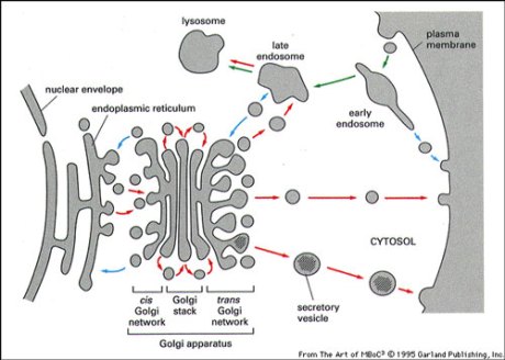
The specialized digestive enzymes and membrane proteins of the lysosomes are synthesized in the ER and transported to the Golgi apparatus to the trans-Golgi network.

Ubiquitin is a protein signal for endosome sorting of membrane proteins which leads the cargo to the lysosome for degradation.

Transport vesicles mediate through two pathways:

  1. Outward Secretory Pathway (goes out through the cell surface); normally includes translocation across the ER followed by transfer to stacked Golgi cisternae (interconnected network of membranes also known as cis-Golgi), then to the trans-Golgi network where the protein will bud-off in a transport vesicle created by the trans-Golgi membrane, a process called exocytosis. 

Outward Secretory Pathway. Click on image for credit.

Cis and trans refers to the different faces of the Golgi complex. Vesicles come into the cis face from the RER and leave from the trans face to the plasma membrane or lysosomes.

Each molecule that travels along this route passes through a fixed sequence of modifications in the membrane and lumen of the RER and Golgi complex;

  • Formation of disulfide bonds– Disulfide bonds (composed of two sulfur atoms united to carbon atoms. More of in biochemistry) help stabilize the structure of proteins. They do not form in the cytosol because of the reducing (electron giving and therefore disrupting the bond) environment there that interrupts the formation of disulfide bonds.
  • Addition and processing of carbohydrates (glycosylation as in –ation or addition of glycose)- Glycosylation can protect the protein from degradation, and also to hold it in the ER until it is properly folded, or help guide it to the appropriate organelle by serving as a transport signal in the transport vesicle (because it can function in recognition).

Example of an N-Linked Glycosylation. Click on image for credit.

Oligosaccharides (14 sugars) side chains link to an amino acid’s (usually asparagine) amide (-NH2) group, specifically to the nitrogen atom, so it is called N-linked glycosylation.

Modifications to N-linked oligosaccharides are completed in the Golgi complex. The middle region of Golgi apparatus contains enzymes- glycosyl transferases– for this purpose. Some proteins are glycosylated by O-linked oligosaccharides (1-4 sugars) in both the ER and Golgi apparatus.

  • Proper folding– several strategies are used to ensure that only proteins properly folded are transported. Some examples are stress response in the ER are ER associated protein degradation (ERAD) and unfolded protein response (UPR).
  • Specific proteolytic cleavages– assembly into multimeric (different polypeptide chains come together) proteins.

         2. Inward Endocytic Pathway (directed to endosomes or lysosomes within the cell);  Endocytosis is a process by which cells take up and absorb molecules into it from the extracellular matrix (ECM) by engulfing them. Part of the plasma membrane is invaginated during this process and pinches of forming a membrane-bound vesicle called an endosome.

Endocytosis. Click on image for credit.

The endosome exists in two phases: an early endosome whose task is to physically separate or sort receptor from ligands (signals) that have been internalized, this is why early endosomes are called sorting endosomes. The next phase is the late endosome involved in the breakdown of internalized cargo.

The two types of endosomes. Early(Sorting) and late endosomes. Click on image for credit.

There are three forms of endocytosis:

Example of Receptor-Mediated Endocytosis (RME). Click on image for credit.

Receptor sorting is the major form of endosome sorting because the receptors engage in rapid recycling. The other two are clear enough without illustrations I guess. If not, then google them 🙂

3- Transport by vesicles: when proteins are made on the rough endoplasmic reticulum (RER), they get loaded into the Golgi apparatus. They are then sorted, modified and packaged in vesicles made from the budding-off of the Golgi membrane and discharged.

Sorting signals directs the protein to the organelle. The signal is usually a stretch of amino acid sequence of about 15-60 amino acids long.

There are at least three principles that characterize all vesicles mediated transport within cells:

                      i.        The formation of membrane vesicles from a larger membrane occurs through the assistance of a protein coat such as clathrin that engulfs the protein because an adapter protein such as adaptin binds both to the coat and to the cargo protein bringing both close together.

Clathrin-coated vesicle transport. Click on image for credit.

The adaptin traps the cargo protein by biding with it’s receptors. After assembly particles bind to the clathrin protein they assemble into a basket-like network on the cytosolic surface of the membrane to shape it into a vesicle. Their final budding-off requires a GTP-binding protein called dynamin.

                    ii.        The process is facilitated by a number of GTP-binding proteins (ex; dynamin) that assemble a ring around the neck of a vesicle and through the hydrolysis of the phosphate group of GTP to GDP until the vesicle pinches off. In other words, GTP is one of the main sources of cellular energy for vesicle movement and fusion.

Dynamin powered by GTP pinches the vesicle off the membrane. Click on image for credit.

 OR

Another picture of the same process. Click on image for credit.

                   iii.        After a transport vesicle buds-off from the membrane, it is actively transported by motor proteins that move along cytoskeleton fibers to its destination. The vesicle then fuses with a target membrane and unloads the cargo (protein). But in order to fuse a vesicle with the membrane of another compartment, they both require complementary proteins, which in this case is soluble N-ethylmalei mide-sensitive-factor attachment protein receptor or, ahem, SNARE present in the membrane – one for the vesicle (vesicular SNARE) and one for the target membrane (t-SNARE).

SNARE helping direct transport vesicles to their target molecules. Click on image for credit.

Each organelle and each type of transport vesicle is believed to carry a unique SNARE. Interactions between complementary SNAREs helps ensure that transport vesicles fuse only with the correct membrane.

Membrane fusing does not always follow immediately after docking (unloading of cargo), however it often waits for specific molecular signals.

Btw, based on coat protein, transport proteins can be classified into three types:


Coated Vesicle Types.

COP stands for Coatamer proteins (COP) which is the second class of coat protein that mediates budding-off of vesicles from large membranes. It can be of two types I (anterograde; moving in a forward direction. Ex; from ER to Golgi) & II (retrograde; moving in a backward direction. Ex: from Golgi back to ER).

2-  Transport across membranes: Similar to how transport functions via the nucleus, most proteins are transported across the biomembrane with the help of receptor proteins which form a complex with the import molecule.

Protein translocators located between the biomembrane (transmembrane protein or integrins) identifies the signal sequence by unfolding the protein and transports it across the membrane into the organelle or cell (similar to the way NPCs do).

Once the protein enters the organelle or cell, its signal sequence is cut off by a peptidase enzyme and the protein is folded into its final 3-D shape again with the help of helper molecules.


In the case of proteins intended for secretion out of the cell, as soon as their N-terminal contains the signal sequence that will indicate this, a signal recognition particle binds to it and forms a protein complex causing synthesis to slow down. The SRP-protein complex will travel and bind to a SRP-receptor on a rough endoplasmic reticulum (RER) membrane. Once the protein gets to the RER membrane, its synthesis will continue at the normal rate until it is complete. During translocation out of the cell, the signal sequence is cut off by a signal peptidase located in the RER membrane and released from the translocation channel and degraded to amino acids.

RER serves as an entering point for proteins that have to go to other organelles like the Golgi complex and lysosome. They can also ferry proteins out of the cell by transport vesicles formed from the budding off of their biomembrane.


There are three mechanisms in which newly synthesized proteins are inserted into the RER membrane (the mechanisms vary with the type of protein);

  • Type I; signal sequence on N-terminals enters first and continues to elongate until a hydrophobic stop-sequence is reached, and then inserted in the membrane and forms the anchor for that protein. Of course then signal sequence is cut by protease.
  • Type II; these proteins have rather long hydrophobic regions that will be anchored in the membrane with the C-terminal leading. Protein continues to be inserted until it reaches the hydrophobic stop-sequence, but the signal sequence is not cut.
  • Type III; same as type I, the only difference is that the signal sequence is not cut.


And according to the number of times that protein passes through the membrane, the proteins of the ER membrane can be divided into single pass transmembrane protein, double pass transmembrane protein, and multi-pass transmembrane protein.


I understand the information here may appear bland. This is because it has been generalized for most process within the cell and cell biologists study the various pathways that exist which can be very overwhelming. Here a brief overview is being discussed such that when you choose to do more detailed study, this generic pattern would have provided you with a basic background.

When protein sorting occurs, they are imported into different places via one of the following three ways:

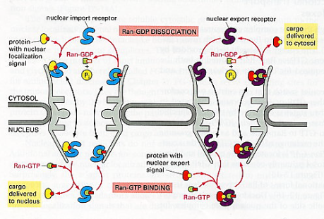
1-    Transport through nuclear pores: Nuclear pores are large complex structures in the nuclear envelope (double-membrane) that regulate movement of macromolecules (proteins and nucleic acids) in and out of the nucleus.

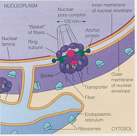
The nuclear envelope has many nuclear pores termed nuclear pore complex (NPC).

Nuclear Pore Complex

In the nucleoplasmic face of the NPC, a nuclear ring supports eight basket filaments joined by the terminal ring forming a structure called the nuclear basket. In the cytoplasmic face, the NPC’s cytoplasmic ring supports eight cytoplasmic filaments. In the center of the NPC, there are the central plug or transporter (protein) and spoke which connect the two ring subunits together.

Nuclear Pore Complex2. Click on image for credit.

This complex provides the nucleus with the ability to selectively allow entry and exit of molecules. Ions, metabolites, and small proteins can pass freely and nonselectively between the nucleus and the cytosol.

Large molecules, however, and macromolecular complexes carry sorting signals called nuclear localization signals (NLS) to which a nuclear import receptor protein (NIRP) will bind and direct through the pore. The NIRP are also called nuclear transport receptors (NTR) and leads the proteins (synthesized in the cytoplasm but required for the nucleus) into the NPC,

The transport process via NPCs.

The NIR attaches to the NLS of the protein that wants to enter the nucleus, forming a NIR-protein complex, and delivers it in through the NPC.

Once inside, one of the proteins of the NPC is an enzyme named Ran-GTPase which functions to add the protein complex Ran-GTP to the NIR-protein complex and causes conformational changes in the latter’s polypeptide chain leading to the dissociation of the NIR from the protein intended for the nucleus. Ran-GTP is a complex of Ran protein attached to guanosine triphosphate (a nucleoside).

The NIR-Ran-GTP complex formed, then, is powered by GTP hydrolysis to GDP (guanosine diphosphate– di meaning two, as in only two phosphate groups remain attached to the guanosine nucleoside molecule) and exit the nucleus through the NPC. The Ran-GTP is thus converted to Ran-GDP which, once the NIR-Ran-GDP complex exits the nucleus, will dissociate from the NIR.

Nuclear export works similar to nuclear import, but in reverse and proteins contain a nuclear-export signal (NES).

Two important concepts will be discussed in this chapter. One is that of the membrane-enclosed organelles which are thought to have evolved in at least two ways:

  1. The nuclear membrane and the membrane of the endoplasmic reticulum, Golgi apparatus, endosomes, and lysosomes are believed to have originated by invagination (inner folding) of the plasma membrane. These membranes and the organelles they enclose (except for the nucleus) are all part of the endomembrane system.

    Simplified illustration of invagination.

  2. Mitochondria and chloroplasts are unique because they have their own DNA that loops in a circle like that of bacteria. And they manufacture many of their own proteins and reproduce by binary fission so they may have evolved in the sense that they were prokaryotes (primordial single-cells) that became so dependent on their host that they are now a part of the cell.


The other important concept is the fact that a typical mammalian cell contains about 10,000 different kinds of proteins. For a cell to work properly, each of these many proteins must be sent to the correct place. The process of directing each newly made polypeptide to particular destination is known as protein targeting or protein sorting.

The proper organelle destination for a protein is determined by signals in the amino acid sequence that will ultimately bind to a receptor protein involved in importing that protein into the organelle.

In some cases, there is a single sequence at the N-terminal which are called signal sequences or signal peptide. And in other cases there are one or more internal sequences that are not in a continuous series called signal patch. Because this sequence is composed of two separate elements, it is referred to as bipartite (being or having two-bi parts-partite).

Different types of signal sequences.

Signal patch are formed because when the protein is made, it is folded and since a fold can bring different regions together (think of smudged paper that can bring the corners of the paper close to the center), the juxtaposition of amino acids from far regions that are physically separated before the protein folds will contain the signal in series (now imagine colouring the smudged paper. After unfolding it, the paper will appear to be coloured in patches). Btw, protein can be synthesized inside the cell or come from outside, and in those two cases it will still have a sequence.

All for now.

Extracellular matrix (ECM) is the material found around cells and biochemically partly made by the cell. It is composed of complex mixtures of proteins, proteoglycans, and, in the case of bone tissue, contains mineral deposits as well.

  • Protein; almost all of the proteins found in the ECM are glycoproteins which include any of a group of complex compounds containing sugar units or polysaccharides combined with amino acid units or polypeptides. Some examples are collagen, laminin and fibronectin. Elastin is not a glycoprotein but is simply a protein and provides flexibility to skin arteries and bones.
  • Proteoglycan; are partially similar to glycoproteins but are made up mostly of carbohydrate consisting of various polysaccharide side chains linked to a protein, and resemble polysaccharides rather than proteins in their properties. They usually have groups of carbohydrate chains attached to a protein backbone.

    Typical Proteoglycan. Click on image for credit.

  • Mineral deposits; found in bones such as calcium and phosphorous.

Most normal cells cannot survive unless they are anchored to the extracellular matrix.

Cells attach to the ECM by means of integrin (integral membrane proteins) which are transmembrane glycoproteins that bind to the ECM proteins collagen, laminin and fibronectin (can exert a physical force on the cell). Internally, the integrin binds to actin filaments of the cytoskeleton. This gives them a key structural role in cells (more on this in chapter 6).


Cells don’t simply squeeze next to each other in a tissue because if they did, they might temporarily hold together but wouldn’t be stable enough to compose tissue as they’d fall out due to lack of adhesion. They are connected to each other via membrane-associated structures (structures situated on the plasma membrane), usually transmembrane proteins. These membrane-associated structures help in cohesion and communication. Where cells stick tightly to each other (adhesion), this is done with the help of Cadherins, a family of transmembrane glycoproteins. Not only does it stick cells together, it also helps seal junctions between cells to prevent the flow of materials through the intercellular space.

There are many types of intercellular junctions situated at various spots on the cell:

1. Tight Junctions or Zonulae Occludens (singular Zonula) situated apically. Zonula is Latin for encircling and Occludensto enclose- so referring to the fact that the junction forms a band completely around the cell fusing cells together and closing-off intercellular space. The number of fusion sites (viewed as ridges and grooves like a suture) often correlate to the leakiness of the cell tissue. The less fusion sites, the more permeable to water and solutes and vice versa. Ex; Cells of the proximal renal tubule of the kidney where filtration often takes place have few fusion sites to allow for leakability, whereas, cells of the urinary bladder (that must hold urine and not allow it to leak out) contains numerous fusion sites or tight junctions to form a seal to prevent the flow of materials between epithelial cells.

Intercellular Junctions. . The cell in the middle was emptied of it’s contents to show the inner surface of it’s membranes.


2. Intermediate Junction or Zonulae Adherens situated at close proximity to the apex but somewhat below the tight junctions, also encircles the cell and provides adhesion of neighboring cells to each other due to the insertion of numerous actin filaments from the cytoplasm of the cell into the junction forming a network. But, unlike tight junctions, they are relatively permeable as there are no fusion sites that completely seal off the paracellular space. The two Zonulas form a continuous ribbon around the cell apex.


3. Gap junction situated almost anywhere along the lateral surface of the epithelial cell is simply a gap between the neighboring cell membranes that is about 2nm. Contains protein unit called connexin (a hexamer: polymer made of six monomers) with a hydrophilic pore (1.5nm diameter) that acts as a hydrophilic ion channel between two cells. Thus, an individual unit of the gap junction is called a connexon (many seen in image).

Connexons. Gap Junction.


4. Desmosome or Macula Adherens can be situated anywhere on the lateral surface (or sides) as well. Macula means a patch so it doesn’t take the length of the membrane and unlike the Zonulas, does not surround the cell. It is a disk-shaped structure matched with an identical structure at the surface of the adjacent cell made up of 12 proteins (circular attachment plaques or spots). The membrane in this region are straight and usually further apart (30nm). It’s adhesion function is further empowered by groups of intermediate keratin filaments which provides firm adhesion.


5. Hemidesmosome literally means half a desmosome. It is also a Macula or plaque or spot of adhesion but between the basal surface of cells and their basal lamina (layer made of connective tissue usually present under cells in tissue). So in effect, the Maculais only on one side.

Intercellular Junctions.


From a functional point of view, therefore, junctions between cells can be classified as adhering junctions (zonula adherens, desmosome and hemidesmosome), impermeable junctions (zonula occludens) and communicating junctions(gap junctions).


Intercellular Junctions Summary Classification.


Additionally, as mentioned earlier, intergrins are receptor proteins that both bind to and respond to the ECM in different ways such as wound healing, cell differentiation (chapter 10), and apoptosis (chapter 11).