Archives for category: proteins

3- Transport by vesicles: when proteins are made on the rough endoplasmic reticulum (RER), they get loaded into the Golgi apparatus. They are then sorted, modified and packaged in vesicles made from the budding-off of the Golgi membrane and discharged.

Sorting signals directs the protein to the organelle. The signal is usually a stretch of amino acid sequence of about 15-60 amino acids long.

There are at least three principles that characterize all vesicles mediated transport within cells:

                      i.        The formation of membrane vesicles from a larger membrane occurs through the assistance of a protein coat such as clathrin that engulfs the protein because an adapter protein such as adaptin binds both to the coat and to the cargo protein bringing both close together.

Clathrin-coated vesicle transport. Click on image for credit.

The adaptin traps the cargo protein by biding with it’s receptors. After assembly particles bind to the clathrin protein they assemble into a basket-like network on the cytosolic surface of the membrane to shape it into a vesicle. Their final budding-off requires a GTP-binding protein called dynamin.

                    ii.        The process is facilitated by a number of GTP-binding proteins (ex; dynamin) that assemble a ring around the neck of a vesicle and through the hydrolysis of the phosphate group of GTP to GDP until the vesicle pinches off. In other words, GTP is one of the main sources of cellular energy for vesicle movement and fusion.

Dynamin powered by GTP pinches the vesicle off the membrane. Click on image for credit.

 OR

Another picture of the same process. Click on image for credit.

                   iii.        After a transport vesicle buds-off from the membrane, it is actively transported by motor proteins that move along cytoskeleton fibers to its destination. The vesicle then fuses with a target membrane and unloads the cargo (protein). But in order to fuse a vesicle with the membrane of another compartment, they both require complementary proteins, which in this case is soluble N-ethylmalei mide-sensitive-factor attachment protein receptor or, ahem, SNARE present in the membrane – one for the vesicle (vesicular SNARE) and one for the target membrane (t-SNARE).

SNARE helping direct transport vesicles to their target molecules. Click on image for credit.

Each organelle and each type of transport vesicle is believed to carry a unique SNARE. Interactions between complementary SNAREs helps ensure that transport vesicles fuse only with the correct membrane.

Membrane fusing does not always follow immediately after docking (unloading of cargo), however it often waits for specific molecular signals.

Btw, based on coat protein, transport proteins can be classified into three types:


Coated Vesicle Types.

COP stands for Coatamer proteins (COP) which is the second class of coat protein that mediates budding-off of vesicles from large membranes. It can be of two types I (anterograde; moving in a forward direction. Ex; from ER to Golgi) & II (retrograde; moving in a backward direction. Ex: from Golgi back to ER).

When protein sorting occurs, they are imported into different places via one of the following three ways:

1-    Transport through nuclear pores: Nuclear pores are large complex structures in the nuclear envelope (double-membrane) that regulate movement of macromolecules (proteins and nucleic acids) in and out of the nucleus.

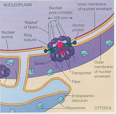
The nuclear envelope has many nuclear pores termed nuclear pore complex (NPC).

Nuclear Pore Complex

In the nucleoplasmic face of the NPC, a nuclear ring supports eight basket filaments joined by the terminal ring forming a structure called the nuclear basket. In the cytoplasmic face, the NPC’s cytoplasmic ring supports eight cytoplasmic filaments. In the center of the NPC, there are the central plug or transporter (protein) and spoke which connect the two ring subunits together.

Nuclear Pore Complex2. Click on image for credit.

This complex provides the nucleus with the ability to selectively allow entry and exit of molecules. Ions, metabolites, and small proteins can pass freely and nonselectively between the nucleus and the cytosol.

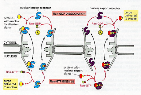
Large molecules, however, and macromolecular complexes carry sorting signals called nuclear localization signals (NLS) to which a nuclear import receptor protein (NIRP) will bind and direct through the pore. The NIRP are also called nuclear transport receptors (NTR) and leads the proteins (synthesized in the cytoplasm but required for the nucleus) into the NPC,

The transport process via NPCs.

The NIR attaches to the NLS of the protein that wants to enter the nucleus, forming a NIR-protein complex, and delivers it in through the NPC.

Once inside, one of the proteins of the NPC is an enzyme named Ran-GTPase which functions to add the protein complex Ran-GTP to the NIR-protein complex and causes conformational changes in the latter’s polypeptide chain leading to the dissociation of the NIR from the protein intended for the nucleus. Ran-GTP is a complex of Ran protein attached to guanosine triphosphate (a nucleoside).

The NIR-Ran-GTP complex formed, then, is powered by GTP hydrolysis to GDP (guanosine diphosphate– di meaning two, as in only two phosphate groups remain attached to the guanosine nucleoside molecule) and exit the nucleus through the NPC. The Ran-GTP is thus converted to Ran-GDP which, once the NIR-Ran-GDP complex exits the nucleus, will dissociate from the NIR.

Nuclear export works similar to nuclear import, but in reverse and proteins contain a nuclear-export signal (NES).

Two important concepts will be discussed in this chapter. One is that of the membrane-enclosed organelles which are thought to have evolved in at least two ways:

  1. The nuclear membrane and the membrane of the endoplasmic reticulum, Golgi apparatus, endosomes, and lysosomes are believed to have originated by invagination (inner folding) of the plasma membrane. These membranes and the organelles they enclose (except for the nucleus) are all part of the endomembrane system.

    Simplified illustration of invagination.

  2. Mitochondria and chloroplasts are unique because they have their own DNA that loops in a circle like that of bacteria. And they manufacture many of their own proteins and reproduce by binary fission so they may have evolved in the sense that they were prokaryotes (primordial single-cells) that became so dependent on their host that they are now a part of the cell.


The other important concept is the fact that a typical mammalian cell contains about 10,000 different kinds of proteins. For a cell to work properly, each of these many proteins must be sent to the correct place. The process of directing each newly made polypeptide to particular destination is known as protein targeting or protein sorting.

The proper organelle destination for a protein is determined by signals in the amino acid sequence that will ultimately bind to a receptor protein involved in importing that protein into the organelle.

In some cases, there is a single sequence at the N-terminal which are called signal sequences or signal peptide. And in other cases there are one or more internal sequences that are not in a continuous series called signal patch. Because this sequence is composed of two separate elements, it is referred to as bipartite (being or having two-bi parts-partite).

Different types of signal sequences.

Signal patch are formed because when the protein is made, it is folded and since a fold can bring different regions together (think of smudged paper that can bring the corners of the paper close to the center), the juxtaposition of amino acids from far regions that are physically separated before the protein folds will contain the signal in series (now imagine colouring the smudged paper. After unfolding it, the paper will appear to be coloured in patches). Btw, protein can be synthesized inside the cell or come from outside, and in those two cases it will still have a sequence.

All for now.

Extracellular matrix (ECM) is the material found around cells and biochemically partly made by the cell. It is composed of complex mixtures of proteins, proteoglycans, and, in the case of bone tissue, contains mineral deposits as well.

  • Protein; almost all of the proteins found in the ECM are glycoproteins which include any of a group of complex compounds containing sugar units or polysaccharides combined with amino acid units or polypeptides. Some examples are collagen, laminin and fibronectin. Elastin is not a glycoprotein but is simply a protein and provides flexibility to skin arteries and bones.
  • Proteoglycan; are partially similar to glycoproteins but are made up mostly of carbohydrate consisting of various polysaccharide side chains linked to a protein, and resemble polysaccharides rather than proteins in their properties. They usually have groups of carbohydrate chains attached to a protein backbone.

    Typical Proteoglycan. Click on image for credit.

  • Mineral deposits; found in bones such as calcium and phosphorous.

Most normal cells cannot survive unless they are anchored to the extracellular matrix.

Cells attach to the ECM by means of integrin (integral membrane proteins) which are transmembrane glycoproteins that bind to the ECM proteins collagen, laminin and fibronectin (can exert a physical force on the cell). Internally, the integrin binds to actin filaments of the cytoskeleton. This gives them a key structural role in cells (more on this in chapter 6).


Cells don’t simply squeeze next to each other in a tissue because if they did, they might temporarily hold together but wouldn’t be stable enough to compose tissue as they’d fall out due to lack of adhesion. They are connected to each other via membrane-associated structures (structures situated on the plasma membrane), usually transmembrane proteins. These membrane-associated structures help in cohesion and communication. Where cells stick tightly to each other (adhesion), this is done with the help of Cadherins, a family of transmembrane glycoproteins. Not only does it stick cells together, it also helps seal junctions between cells to prevent the flow of materials through the intercellular space.

There are many types of intercellular junctions situated at various spots on the cell:

1. Tight Junctions or Zonulae Occludens (singular Zonula) situated apically. Zonula is Latin for encircling and Occludensto enclose- so referring to the fact that the junction forms a band completely around the cell fusing cells together and closing-off intercellular space. The number of fusion sites (viewed as ridges and grooves like a suture) often correlate to the leakiness of the cell tissue. The less fusion sites, the more permeable to water and solutes and vice versa. Ex; Cells of the proximal renal tubule of the kidney where filtration often takes place have few fusion sites to allow for leakability, whereas, cells of the urinary bladder (that must hold urine and not allow it to leak out) contains numerous fusion sites or tight junctions to form a seal to prevent the flow of materials between epithelial cells.

Intercellular Junctions. . The cell in the middle was emptied of it’s contents to show the inner surface of it’s membranes.


2. Intermediate Junction or Zonulae Adherens situated at close proximity to the apex but somewhat below the tight junctions, also encircles the cell and provides adhesion of neighboring cells to each other due to the insertion of numerous actin filaments from the cytoplasm of the cell into the junction forming a network. But, unlike tight junctions, they are relatively permeable as there are no fusion sites that completely seal off the paracellular space. The two Zonulas form a continuous ribbon around the cell apex.


3. Gap junction situated almost anywhere along the lateral surface of the epithelial cell is simply a gap between the neighboring cell membranes that is about 2nm. Contains protein unit called connexin (a hexamer: polymer made of six monomers) with a hydrophilic pore (1.5nm diameter) that acts as a hydrophilic ion channel between two cells. Thus, an individual unit of the gap junction is called a connexon (many seen in image).

Connexons. Gap Junction.


4. Desmosome or Macula Adherens can be situated anywhere on the lateral surface (or sides) as well. Macula means a patch so it doesn’t take the length of the membrane and unlike the Zonulas, does not surround the cell. It is a disk-shaped structure matched with an identical structure at the surface of the adjacent cell made up of 12 proteins (circular attachment plaques or spots). The membrane in this region are straight and usually further apart (30nm). It’s adhesion function is further empowered by groups of intermediate keratin filaments which provides firm adhesion.


5. Hemidesmosome literally means half a desmosome. It is also a Macula or plaque or spot of adhesion but between the basal surface of cells and their basal lamina (layer made of connective tissue usually present under cells in tissue). So in effect, the Maculais only on one side.

Intercellular Junctions.


From a functional point of view, therefore, junctions between cells can be classified as adhering junctions (zonula adherens, desmosome and hemidesmosome), impermeable junctions (zonula occludens) and communicating junctions(gap junctions).


Intercellular Junctions Summary Classification.


Additionally, as mentioned earlier, intergrins are receptor proteins that both bind to and respond to the ECM in different ways such as wound healing, cell differentiation (chapter 10), and apoptosis (chapter 11).

In terms of permeability, water, dissolved gases such as carbon dioxide and oxygen and lipid solid molecules simply diffuse across the phospholipid bilayer because the fluidity permits them.

Water soluble anions (negative ions) generally pass through small horns less than .8 nm in diameter (.0000000008m; 1m=1000mm, 1mm=1000µ-micrometer, 1µm= 1000nm). However, all other larger molecules require carrier molecules or proteins to transport them through the membrane.


The following are an overview of the four different ways in which atoms and molecules cross the biomembrane, which is also called transmembrane transport:


  • Simple diffusion; is the process by which small molecules and ions simply, because of their tendency to spontaneously move around, especially from a region where they are highly concentrated towards a region where they are less, diffuse through the phospholipid bilayer of the biomembrane.


  • Facilitated diffusion; is the process by which, apart from water and ions, specific molecules such as monosaccharides and amino acids, diffuse through channel proteins down their concentration gradient. This assisted diffusion provides them with a special hydrophilic pathway since some molecules might resist the hydrophobic core of the phospholipid bilayer. Some specific ion channels remain open much of the time and are called nongated channels and others only open in response to specific chemical or electrical signals and referred to as gated channels, including the voltage-gated channels of the nervous system which transmit action potentials (nerve impulses) to the brain.


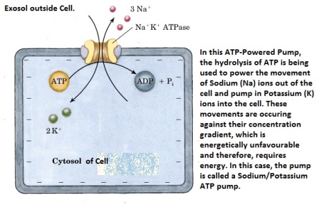
  • Active Transport; is the transmembrane transport that occurs when channel proteins, such as the enzyme ATPases (enzymes usually have the suffix –ase) use the energy of ATP hydrolysis (breaking one phosphate group from the nucleotide Adenosine triphosphate) to move ions or small molecules across a membrane against their chemical concentration gradient or electric potential gradient or both.


General Principle of ATP hydrolysis. Breaking bonds usually give off energy.


ATP-powered pumps which requires energy, is coupled to the hydrolysis of ATP and the overall reaction– ATP hydrolysis and the “uphill” movement of ions or small molecules- is energetically favorable. All ATP-powered pumps are transmembrane proteins with one or more binding sites for ATP located on the cytosolic face of the membrane.

Sodium/Potassium ATP Powered Pump.

Although these proteins commonly are called ATPases, they normally do not hydrolyze ATP into ADP and Pi unless ions or other molecules are simultaneously transported. Because of this tight coupling between ATP hydrolysis and transport, the energy stored in the phosphahydride bond (bond between two phosphate groups) is not dissipated but rather used to move ions or other molecules uphill against an electrochemical gradient.


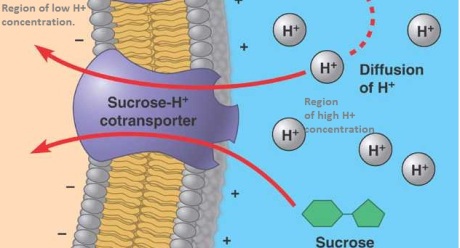
  • Cotransporter; are also enzymes. They mediate coupled reactions in which an energetically unfavorable reaction, such as the movement of molecules or ions against their concentration gradient, is coupled to an energetically favorable reaction such as the passive transport of molecules down their concentration gradient. The co-transporter will use the energy of the passive movement to actively transport other molecules up their concentration gradient. Therefore, unlike ATP, two molecules have to always move through the co-transporter at one time.


This Cotransporter is a Symporter. More below. Click on image for credit.

 The co-transporter is sometimes referred to as secondary-active transporter because it uses the energy stored into an electrochemical gradient.

Cotransporters fall into two types:

1-            Antiporter: in which the movement of both molecules (energy favorable and unfavorable) are in opposite directions.

2-            Symporter: in which the movement of both molecules are in the same direction (see above image). This should not confuse you because the same principle applies- one molecule will be moving down its concentration gradient, and the energy in this will be used to actively move another molecule up its concentration gradient but in the same direction, similar to the Sodium/Potassium pump but without the need for ATP hydrolysis because the Cotransporters are smarter and more efficient than Active transporters or ATPases.

3. Apart from membrane-lipids and membrane-proteins, the final basic component of a biomembrane is membrane-carbohydrates. However, they do not exist in isolation, rather in combination with membrane-lipids to form glucolipids/glycolipids, or combine with membrane-proteins to form glucoproteins/glycoproteins. The combination happens via covalent bonds.

Membrane-Lipids and -Proteins. Click on image for credit.

Most membrane carbohydrates are located on plasma membranes and have the following functions:

  • Improve the stability of membranes.
  • Advance the fastness and activate proteinase (protein enzymes which hydrolyze proteins into polypeptides) in the extracellular matrix.
  • Help membrane-proteins form correct three-dimensional configurations.
  • Take part in cell signal recognition, cell adhesion and cell junction (more detail later).
  • Correct the position and orientation of new proteins for transfer.
  • Lectin, a glucoprotein, protects the cell. So they also aid in protection.
  • Glucolipids act as blood groups determinants because some function as cell recognition particles.

I understand that stating these important roles is not enough to fully comprehend their significance. Later chapters will deal with the details. This is simply a fleeting reference to their roles.

In general, according to their main functions proteins and glycoproteins can be classified to five types:

  1. Channel proteins to form pores for the free transport of small molecules and ions across the membrane (more in Chapter 4).
  2. Carrier proteins to facilitate diffusion and active transport of molecules and ions across the membrane (more in next post).
  3. Cell Recognition Proteins; identifies a particular cell or gives them identity (more in Chapter 7).
  4. Receptor proteins; binds with specific molecules such as hormones and cytokines and gets activated to perform a specific function (also more in chapter 7).
  5. Enzymatic proteins; catalyze specific chemical reactions.
I think it is becoming clear that proteins are really important macro molecules and that most of animal cell biology will discuss it’s various functions.